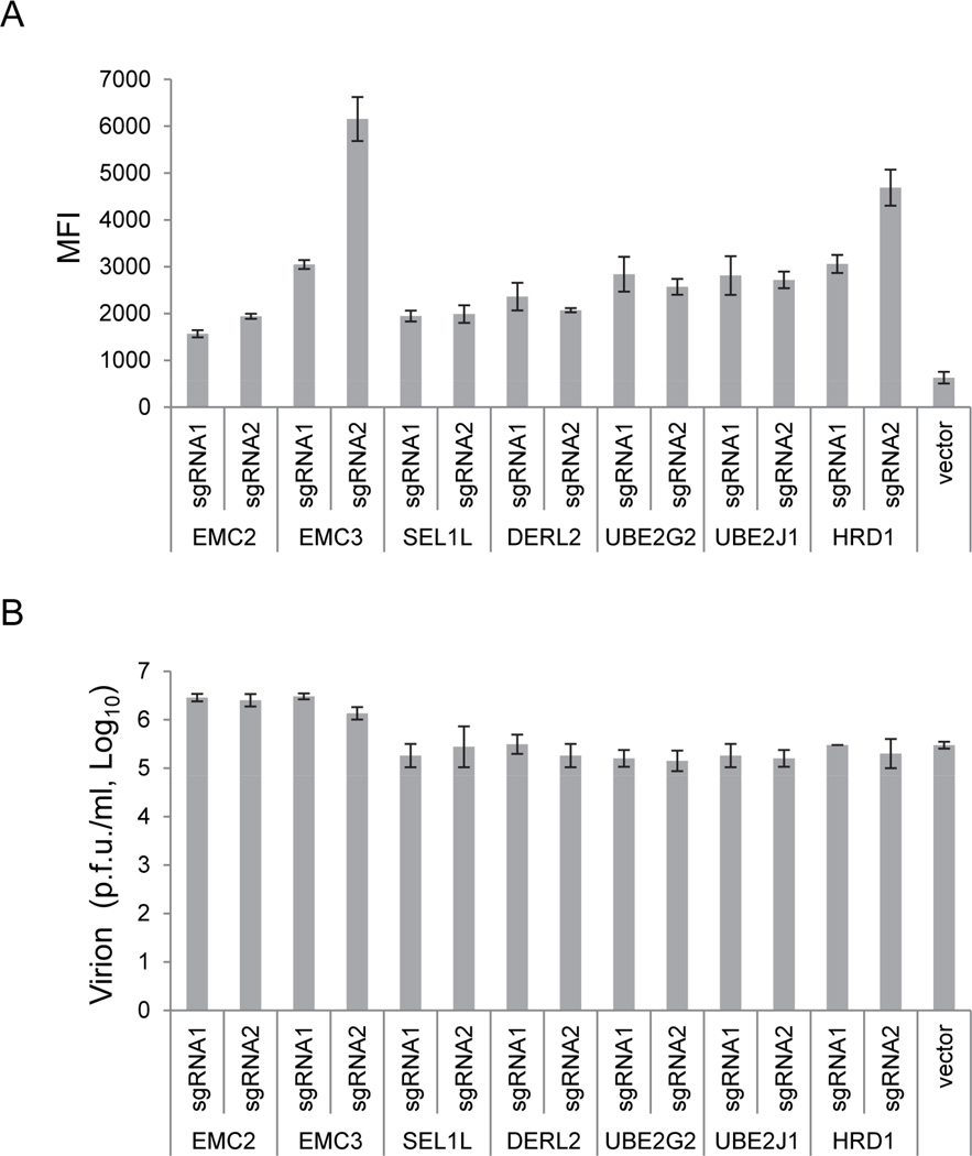
Figure 3. Knockout of the genes (from Figure 2) did not block WNV replication.
(A) Cells derived as in Figure 2A were fixed and stained with WNV envelope (ENV) protein antibody 72 hours after WNV (strain B956) challenge and analyzed by flow cytometry. The mean fluorescence index (MFI) was used to represent the WNV ENV protein level, and see also Figure S2. Vector-transfected cells were included as a control, and the cells were fixed and analyzed at 24 hours post infection, which is at the peak of WNV replication in WT 293FT cells. Error bar, 1 S.D. (n=3). (B) Cells were reseeded into a new 24-well plate. Supernatants were collected 72 hours later, and viral titers were determined by plaque assay. The control reference level was the virus titer in the supernatant of WT cells infected with WNV at 36 hours, which is the peak level of virus in the supernatant. Error bar, 1 S.D. (n=3).

