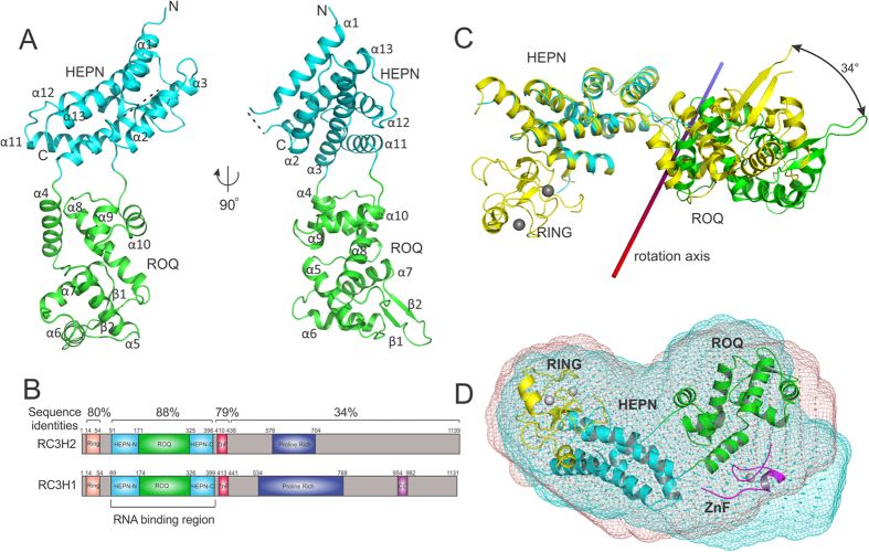
Figure 1. Characterization of the apo-form RC3H2 structure and comparison with RC3H1.
(A) Ribbon diagram of the structure of the RC3H2 RNA-binding region. The ROQ and HEPN domains are coloured in green and cyan respectively. (B) Domain organization of full-length human RC3H1 and RC3H2. (C) Superposition of apo-RC3H2 (PDB:4Z30) and apo-RC3H1 structures (PDB:4TXA). The two structures are aligned in reference to the HEPN domain. The colour scheme for RC3H2 is the same as that in panel (A); RC3H1 is shown in yellow and zinc ions are shown as grey spheres. (D) SAXS envelope models of RC3H1 (a.a. 1–445, cyan) and RC3H2 (a.a. 1–442, red). For model fitting, the HEPN+ROQ domains of RC3H1 (PDB:4TXA) are replaced with the apo-structure of RC3H2, while keeping the orientation between the HEPN(cyan) and RING (yellow) domains unchanged. The ZnF domain (magenta) is built from PDB:1RGO. The structures were fitted to the SAXS envelope of RC3H2.

