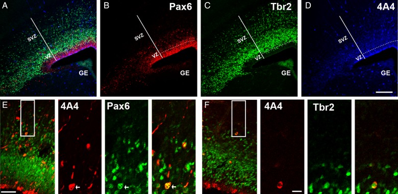
Figure 2.
Analysis of precursor cell number. Maternal MAUab and MTDab IgGs were injected in utero in E14 mice, and precursor cell number in the cerebral cortex was quantified 2 days later at E16. (A) Triple immunostaining for Pax6 (red, B), Tbr2 (green, C), and 4A4 (blue, D). (E) Proliferative precursor cell types located in the SVZ of the E16 developing cortex. Inset indicates region of higher power images to the right. Mitotic translocating RG cells were identified by expression of the mitotic cell marker 4A4 (red), presence of a pial directed fiber, and expression of Pax6 (green). (F) Proliferative precursor cell types in the E16 developing cortex. Inset indicates region of higher power images to the right. Mitotic intermediate progenitor cells were identified by expression of the mitotic cell marker 4A4 (red), and expression of Tbr2 located in the SVZ (green). Calibration bar: (A–D) 250 µm; (E,F) 100 µm and 25 µm, respectively.

