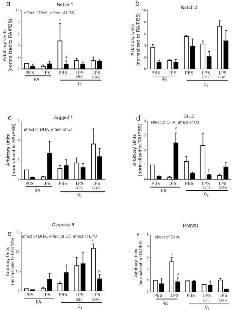
Figure 2.
Western blot analyses for Notch pathway (a–d), caspase 9 (e), and HMGB1 (f) proteins were performed on homogenates from primary macrophages isolated from mice fed standard diets, supplemented with vehicle or DHA in culture (in vitro), and subsequently treated with O2 and/or LPS. Separation was performed by standard protocols as described in Methods and blots were quantified by densitometry. White bars indicate vehicle, black bars indicate DHA supplement. Data were analyzed by using a Multivariate Linear Regression Models with diet as a fixed factor, treatment and exposure as co-variants, and 2 and 3-way interactions were assessed. Differences within individual groups was analyzed by Tukey's post hoc. The data reflect n = 3 from three independent experiments. Major effects and interactions are indicated on the graphs. Post hoc analyses are indicate by * different than CD-RA/PBS; # different than same treatment (difference between diets), p < 0.05.

