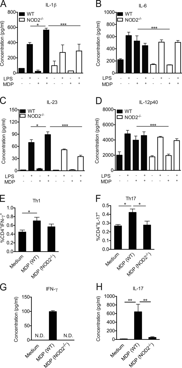
Figure 3.
NOD2 activation triggers a proinflammatory response in vitro. Bone marrow–derived DCs from WT or NOD2−/− mice were stimulated with LPS alone (200 ng/ml) for 24 h in the presence or absence of MDP (10 µg/ml). The levels of IL-1β (A), IL-6 (B), IL-23 (C), and IL-12p40 (D) were measured in the supernatant by ELISA. Alternatively, bone marrow–derived DCs from WT or NOD2−/− mice were co-cultured with naive CD4+ T cells in the presence of MDP (10 µg/ml) for 5 d and were then assessed for the presence of CD4+ IFN-γ+ (Th1; E) and CD4+IL-17+ (Th17; F) cells by flow cytometry. The concentrations of IFN-γ (G) and IL-17 (H) were measured in the supernatant by ELISA. The results are expressed as the mean ± SEM. At least four technical replicates per group were used in each in vitro experiment. *, P ≤ 0.05; **, P ≤ 0.01; ***, P ≤ 0.001.

