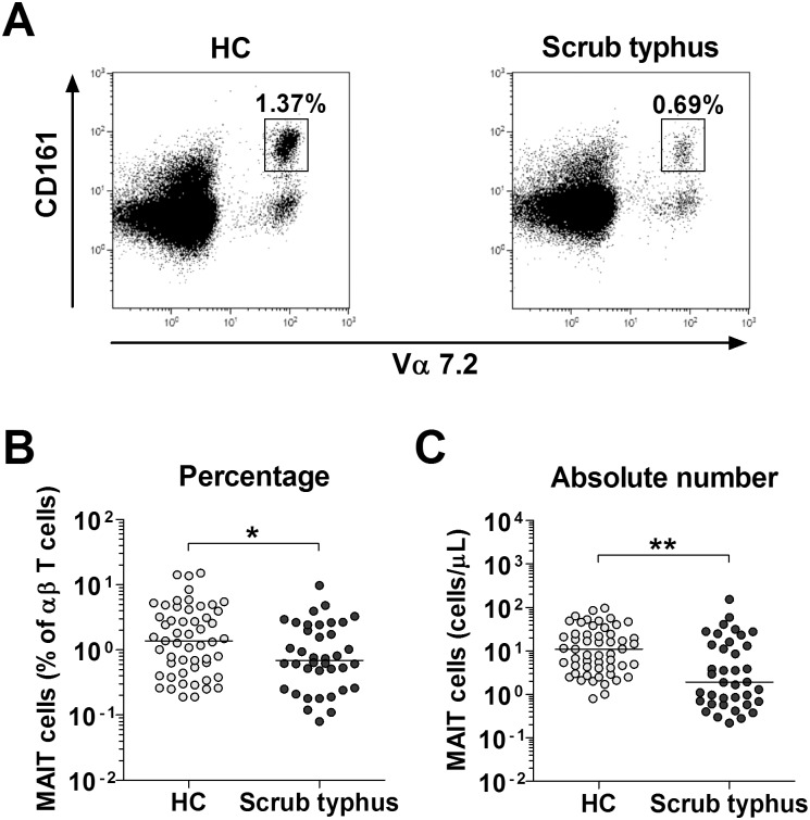
Fig 1. Decreased circulating MAIT cell numbers in the peripheral blood of scrub typhus patients.
Freshly isolated PBMCs from 53 HCs and 38 patients with scrub typhus were stained with APC-Alexa Fluor 750-conjugated anti-CD3, FITC-conjugated anti-TCR γδ, APC-conjugated anti-TCR Vα7.2 and PE-Cy5-conjugated anti-CD161 mAbs, and then analyzed by flow cytometry. Percentages of MAIT cells were calculated using a αβ T cell gate. Panel A: Representative MAIT cell percentages as determined by flow cytometry. Panel B: MAIT cell percentages among peripheral blood αβ T cells. Panel C: Absolute MAIT cell numbers (per microliter of blood). Symbols represent individual subjects and horizontal lines are median values. *p < 0.05, **p < 0.001 by the Mann-Whitney U test.

