Figure 2.
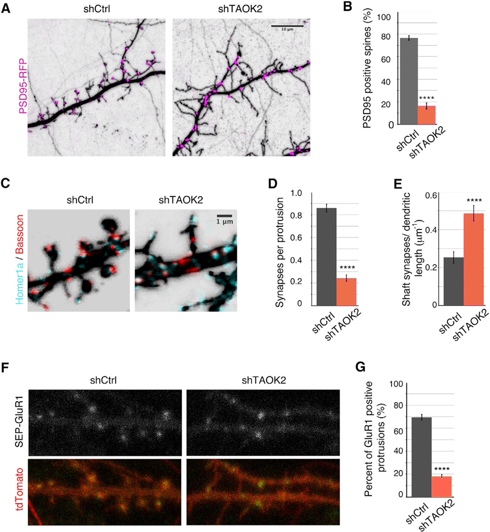
Synapses Are Formed Directly on the Dendritic Shaft in Absence of TAOK2
(A) Hippocampal neurons transfected with either control or TAOK2 shRNA along with RFP-tagged PSD95 to visualize postsynaptic localization of PSD95. The scale bar represents 10 µm.
(B) Percent dendritic protrusions positive for PSD95 (n =15 per condition, p < 0.0001, and t test).
(C) DIV18 neurons transfected with either control or shRNA against TAOK2 were fixed and immunostained for presynaptic Bassoon and postsynaptic Homer1a. The co-localization of Bassoon/Homer1 a in confocal images was considered as a synapse. The scale bar represents 1 µm.
(D) Number of synapses made on the dendritic protrusions as a fraction of total protrusions (n = 10 neurons per condition, p < 0.0001, and t test).
(E) Number of synapses made directly on the dendritic shaft per unit dendritic length (n = 10 neurons per condition, p < 0.0001, and t test). The error bars represent SEM.
(F) Representative confocal images of hippocampal neurons transfected with either control or TAOK2 shRNA (expresses tdTomato under separate promoter) along with pH-sensitive SEP-GluR1. The scale bar represents 5 µm.
(G) Percent of GluR1 positive protrusions (n = 15 per condition, p < 0.0001, and t test). All of the error bars represent SEM.

