
Figure 2. Example brain images indicating the presence of increased extra-axial CSF.
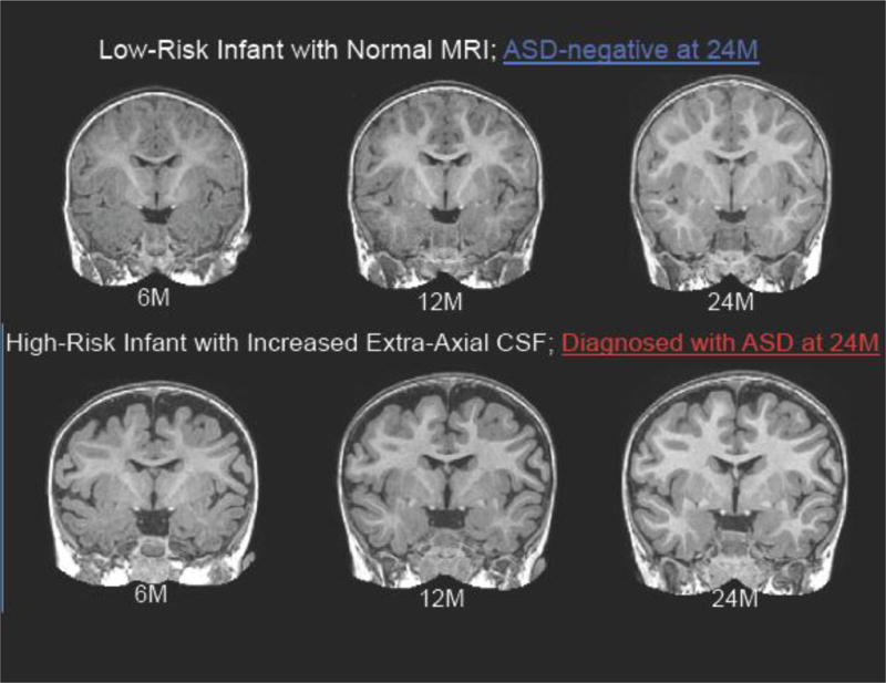
(A) T1-weighted coronal images of a low-risk infant with normal MRI at 6, 12, and 24 months. (B) T1-weighted coronal images of a high-risk infant with increased extra-axial CSF at 6, 12, and 24 months. This child was diagnosed with ASD at 24 months.