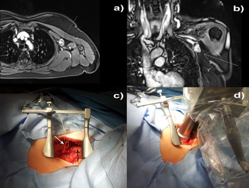
Figure 5.

Intraoperative treatment: (a) axial and (b) coronal magnetic resonance images showing the relationship between the 3 cm irregular auxiliary mass to the auxiliary vessel/nerve complex; (c) the auxiliary vessels exposed (black arrow), thoracodorsal nerve placed aside (white arrow); and (d) the applicator setup.
