
Fig. 2.
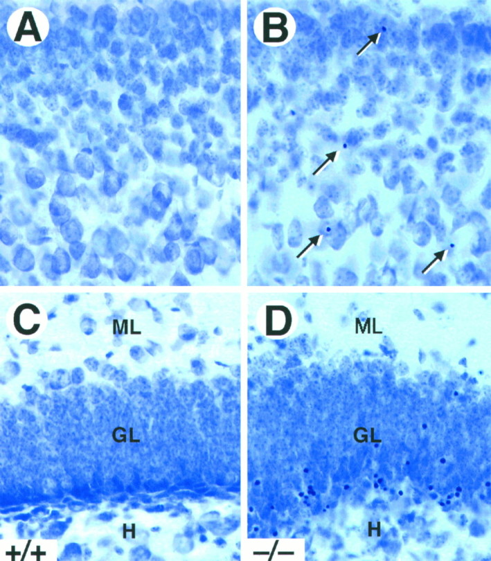
Increased cell death in trkB (−/−) mice. A–D, Cresyl violet-stained sections of the neocortex (A, B) and dentate gyrus (C, D) of P16 control (A, C) andtrkB (−/−) (B, D) mice. Notice the presence of pyknotic nuclei in layers II and III of the neocortex (B, arrows) and the granular layer of the dentate gyrus (D) in the trkB (−/−) mutant mice.GL, Granular layer; H, hilus;ML, molecular layer. Magnification, 40×.