
Fig. 4.
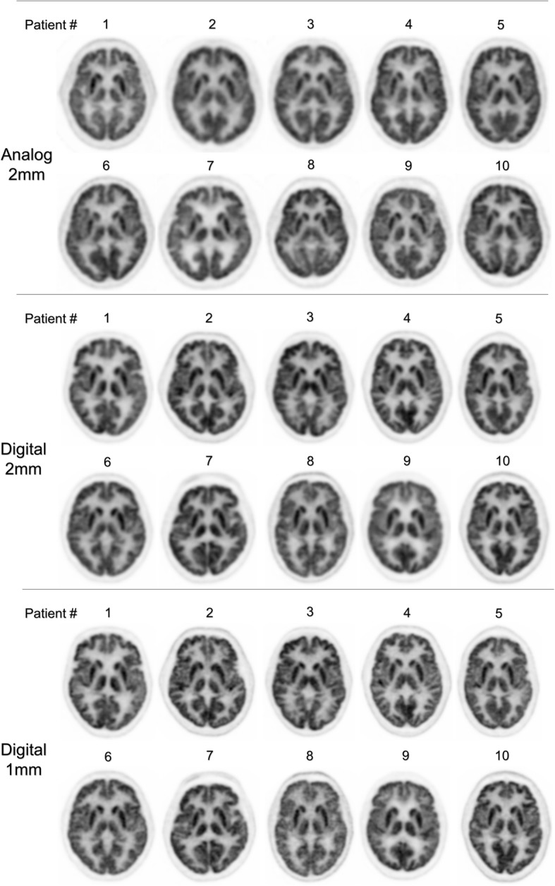
Gallery of axial brain 18F-FDG PET images obtained with analog PET and a current reconstruction method using a 2-mm voxel size (n = 10) (upper row), with digital PET and either a comparable reconstruction method with 2-mm voxel size (n = 10) (middle row) or a high-resolution reconstruction method with 1-mm voxel size (n = 10) (lower row)