
Figure 5.
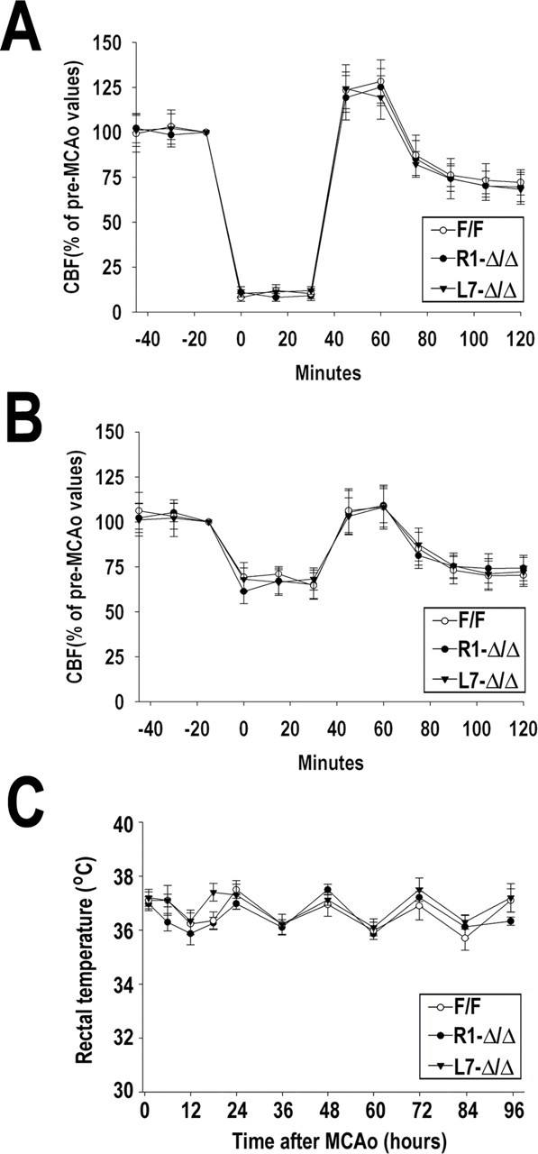
Cerebral blood flow and rectal temperature after MCAo. Local CBF was monitored before, during MCAo, and after reperfusion by laser Doppler flowmetry. Graphs indicate CBF monitored at the ischemic area (A) and the periphery (B). Pre-ischemic values were arbitrarily defined as 100%. Rectal temperature (degrees Celsius) was measured using a rectal probe at 12 h intervals for up to 4 d after ischemia (C). Data are expressed as mean ± SD from 8–10 animals per genotype.