
Figure 2.
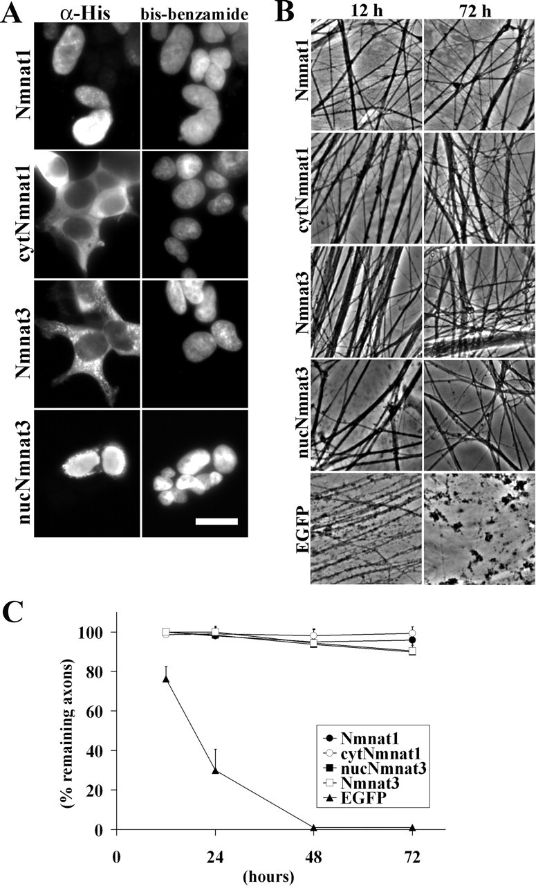
Nmnat enzymes located in the nucleus, cytoplasm, or mitochondria all promote axonal protection. A, Subcellular localization of Nmnat1, cytNmnat1, Nmnat3, or nucNmnat3 in HEK293T cells. Immunohistochemistry using antibody against the hexahistidine epitope tag was used to detect each protein. The cells were also stained with bis-benzimide. As expected, the cytNmnat1 mutant is located in the cytoplasm, and the nucNmnat3 mutant is located in the nucleus. Scale bar, 10 μm. B, In vitro Wallerian degeneration assay using lentivirus-infected DRG neuronal explant cultures expressing Nmnat1, cytNmnat1, Nmnat3, nucNmnat3, or EGFP control. Representative pictures taken at 12 and 72 h after transection show robust protection against axonal degeneration regardless of the subcellular distribution. C, Quantitative analysis of axonal degeneration in DRG explant cultures expressing Nmnat1, cytNmnat1, Nmnat3, or nucNmnat3 at 12, 24, 48, and 72 h after transection. Nmnat1-, cytNmnat1-, Nmnat3-, and nucNmnat3-expressing cells have a significant difference (p < 0.0001; n = 6) with EGFP-expressing cells at 24, 48, and 72 h after axotomy.