
Figure 3.
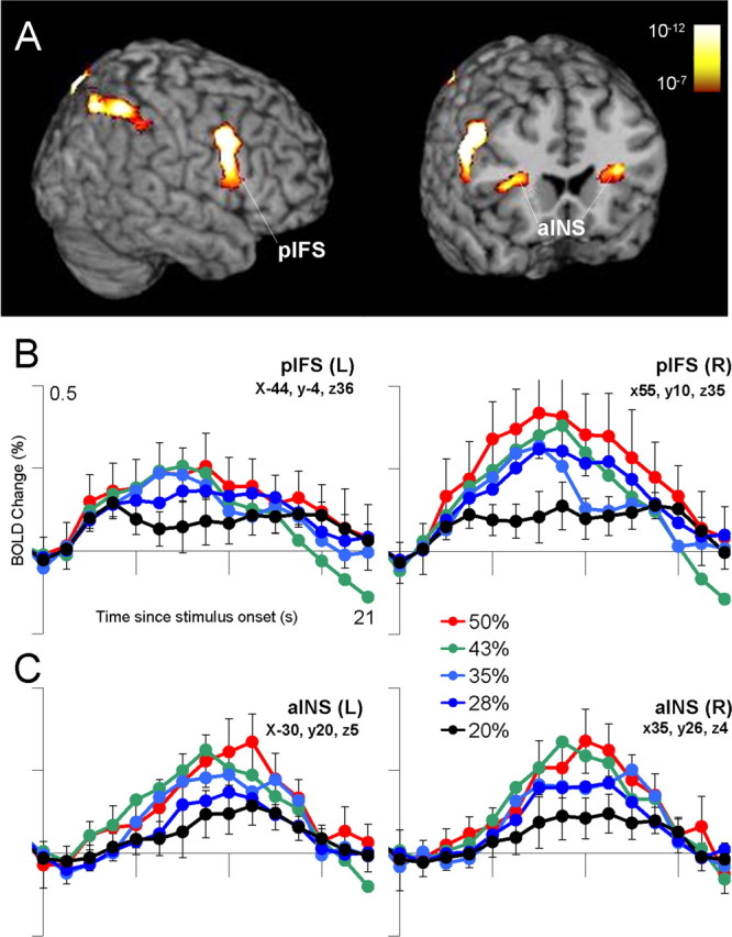
Regions of frontal and insular cortices for which task-related activation was significantly modulated by uncertainty. A, Lateral and cross-sectional views of frontal cortex, with the red-yellow color map (corrected p range, 0.05-0.000001; uncorrected range, 10-7 to 10-12) indicating regions that exhibited significant task-related activation for which amplitude across conditions depended on stimulus uncertainty. B, Within regions of dlPFC, notably the posterior inferior frontal sulcus (pIFS), significant effects of uncertainty on fMRI signal were observed. In these regions, fMRI activation increased as the proportion of uncertainty in the decision increased. L, Left; R, right. C, Similar uncertainty effects were observed within the anterior insula. All of the time courses on this and subsequent figures present amplitude of fMRI BOLD signal (y-axis) as a function of the time since the onset of the context-phase stimuli (x-axis).