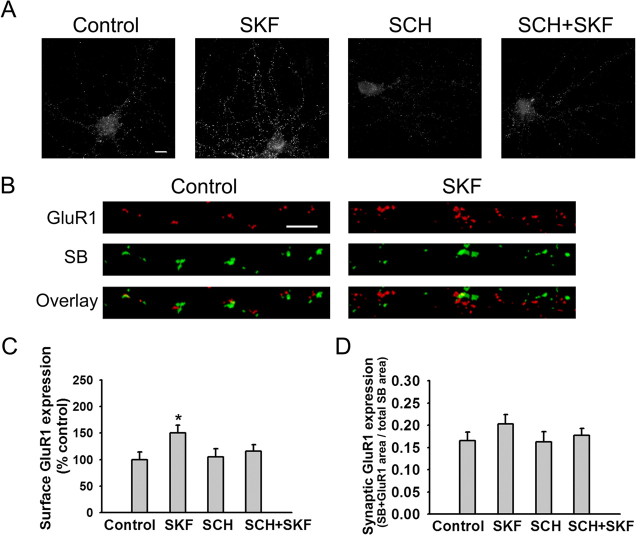
Figure 2.
The D1 receptor agonist SKF 81297 increases GluR1 surface expression but not synaptic incorporation in PFC neurons. A, Representative images of total cell-surface GluR1 staining in control neurons and neurons treated with SKF 81297 (1 μm; 5 min), the D1 receptor antagonist SCH 23390 (10 μm), or SCH plus SKF (SCH+SKF). In the SCH+SKF group, SCH 23390 was added 5 min before SKF. Scale bar, 10 μm. B, Representative images illustrating synaptic GluR1 incorporation, indicated by overlap of GluR1 and SB staining, on processes of control neurons and neurons treated with SKF 81297 (1 μm; 5 min). Scale bar, 5 μm. C, Quantification of total surface GluR1 staining. SKF significantly increased surface GluR1 staining compared with the control group (n = 19-24; Dunn's test; *p < 0.05). Results are presented as the mean area of GluR1 puncta, normalized to the control group. D, Quantification of synaptic GluR1 incorporation, expressed as the fraction of total SB staining area that overlaps with GluR1 staining area, based on analysis of images such as those in B. The SKF, SCH, and SCH+SKF groups were not significantly different from the control group (n = 19-24; ANOVA; p > 0.05). Error bars indicate SEM.

