
Figure 5.
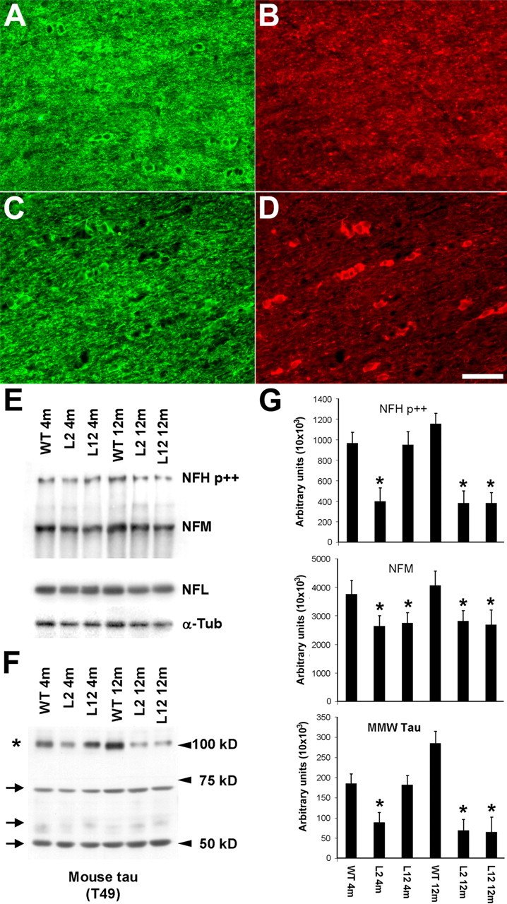
Biochemical impact of OLG tau accumulations on axons in the optic nerve of the PL Tg mice. A, Overexpression of Tg tau in the optic nerve interfascicular OLGs shown by double-immunofluorescence staining with anti-CNP antibody (A, C) and anti-tau antibody 17026 (B, D) for 6-month-old WT non-Tg (A, B) and line 12 PL tau Tg (C, D) mice. In contrast to diffuse tau immunoreactivity in WT OLGs, strong tau staining is seen in specific CNP-positive OLGs of the PL Tg mouse. CNP staining in myelin and tau staining in axons were moderately diminished in the PL Tg mouse compared with the WT mouse. E-G, Immunoblotting for optic nerve samples with antibodies against phosphorylated NFH (NFH p++), NFM, NFL, α-tubulin (α-tub) (E), and endogenous mouse tau (F), and quantification of the blots (n = 4 in each group) (G). Substantial reduction of NFH and NFM occurred in the line 2 and line 12 PL Tg mice at 4 months of age (E and top and middle panels in G). The 12-month-old PL Tg mice also showed slight decrease in the amounts of NFL and α-tubulin. There were no remarkable differences in the levels of low-molecular-weight tau isoforms ranging from 50 to 70 kDa (indicated by arrows) between the WT and PL Tg mice, whereas middle-molecular-weight tau (∼100 kDa, indicated by an asterisk) in the PL Tg mice was pronouncedly reduced relative to the WT mice (F and bottom panel in G). *p < 0.05 by multiple comparison by ANOVA. Scale bar: A-D, 50 μm.