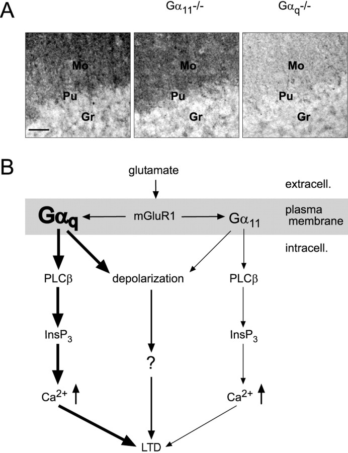
Figure 8.
Gαq as the dominating Gq protein in cerebellar Purkinje cells. A, Immunohistochemistry with an anti-Gαq/Gα11 antibody in cerebellar sections in mice of three genotypes, as indicated. In wild-type and Gα11-deficient mice, the molecular layer, in which the dendritic trees of the Purkinje cells are located, is intensely stained, whereas in Gαq-deficient mice, only very weak staining is present in the molecular layer. Mo, Molecular layer; Pu, Purkinje cell layer; Gr, granule cell layer. Scale bar, 50 μm. B, Model for the mGluR1-dependent signal transduction through Gαq and Gα11 in cerebellar Purkinje cells. Both Gαq and Gα11 are involved in signal transduction pathways leading from activation of mGluR1 to release of Ca2+ from dendritic endoplasmic reticulum and/or to a sEPSP. However, the dominating Gq protein in cerebellar Purkinje cells is Gαq, according to its prevalent expression level compared with Gα11. The mGluR-mediated slow depolarization in Purkinje cells is G-protein dependent.

