
Fig. 1.
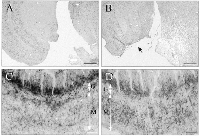
Location and efficacy of ERC lesions implemented in this study. A, B, Photomicrographs of cresyl violet-stained sections through the ERC of Tg mice that underwent sham surgeries (A) or ERC lesions (B). Note the normal anatomy of ERC inA and the extent and specificity of ERC ablation (arrow) in a representative lesion case (B). C, D, AChE-stained sections through the dentate gyrus of Tg mice with sham surgeries (C) or ERC lesions (D). Note the typical change in the AChE staining pattern after ERC lesions, i.e., the diminution of the intense AChE (+) band in the supragranular region and the increased density (sprouting) of AChE fibers toward the outer half of the molecular cell layer as a result of synaptogenesis in septal cholinergic afferents.G, Granule cell layer; M, molecular cell layer. Scale bars: A, B, 400 μm;C, D, 40 μm.