
Fig. 2.
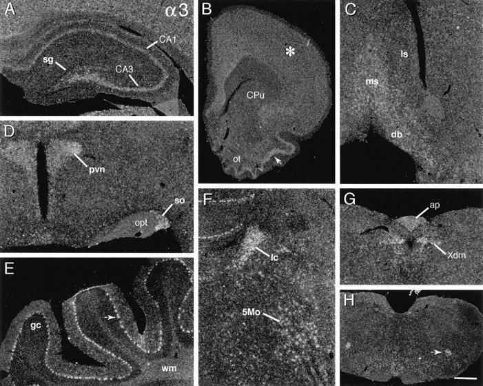
Integrin α3 mRNA is broadly distributed. Dark-field photomicrographs show the autoradiographic localization of α3 cRNA labeling in select brain regions. A, Rostral hippocampal section showing that labeling is distributed within the pyramidal cell layer (CA3, CA1), but not within stratum granulosum (sg). B, Rostral forebrain section shows that labeling is broadly distributed within neocortex (asterisk), moderately dense within superficial piriform cortex (arrow) and olfactory tubercle (ot), but low within caudate putamen (CPu). C, Within the septal region, hybridization is moderately dense in the medial septal nucleus (ms) and the diagonal bands of Broca (db) but is at low neuropil levels within lateral septum (ls). D, Section through hypothalamus showing relatively dense labeling in the paraventricular (pvn) and supraoptic (so) nuclei (opt, optic tract). E, Photomicrograph showing dense labeling of the cerebellar Purkinje cells (arrow) (gc, granule cell layer;wm, deep cerebellar white matter). F, Section showing dense hybridization in the locus coeruleus (lc) and in association with the large neurons of the motor trigeminal nucleus (5Mo). G,H, Sections through lower brainstem showing labeling within the dorsal motor nucleus of the vagus (G;Xdm), area postrema (G;ap), and nucleus ambiguus (H; arrow). Scale bar (shown in H): A, 400 μm; B, 1.2 mm; C, H, 900 μm; D, 320 μm; E, F, 250 μm; G, 450 μm.