
Fig. 8.
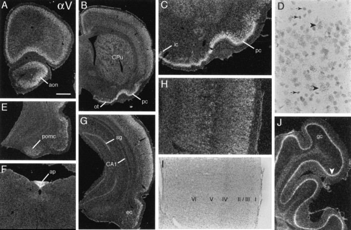
αV mRNA is distributed broadly in cortex and striatum. A, B, Sections through extreme rostral (A) and somewhat more caudal (B) forebrain show broadly distributed αV cRNA labeling in neocortex, anterior olfactory nucleus (aon), caudate putamen (CPu), piriform cortex (pc), and olfactory tubercle (ot).C, Higher magnification view of basal forebrain shows labeling is dense within piriform layer II (pc) and scattered deeper cells and is light in the islands of Calleja (ic). D, High-magnification bright-field photomicrograph of superficial neocortex shows that labeling is associated with larger cells with the Nissl-staining characteristics of neurons (arrowheads) and not with smaller, more densely stained glial-like cells (small arrows).E–G, Sections show hybridization within posteromedial cortical amygdala (pomc; E), area postrema (ap; F), and caudal hippocampus (G). G, Shown is light labeling in hippocampal CA1 stratum pyramidale and stratum granulosum (sg) and labeling in entorhinal cortex (ec). H, I, Photomicrographs of the same field of visual cortex, using dark-field (H) and bright-field (I) illumination to visualize the distribution of αV cRNA labeling (H) relative to cytoarchitectonic lamination (I); labeling is most prominent in layers 2/3 and in association with large cells in deep layer 5, with diffuse and lower density labeling in the nonpyramidal layer 4.J, Field of cerebellar cortex showing rather continuous labeling of the Purkinje cell layer (arrowhead;gc, granule cell layer). Scale bar (shown inA): A, B, 1.0 mm;C, 50 μm; D, 40 μm; E,G, 820 μm; F, 585 μm;H, I, 410 μm; J, 340 μm.