
Figure 2.
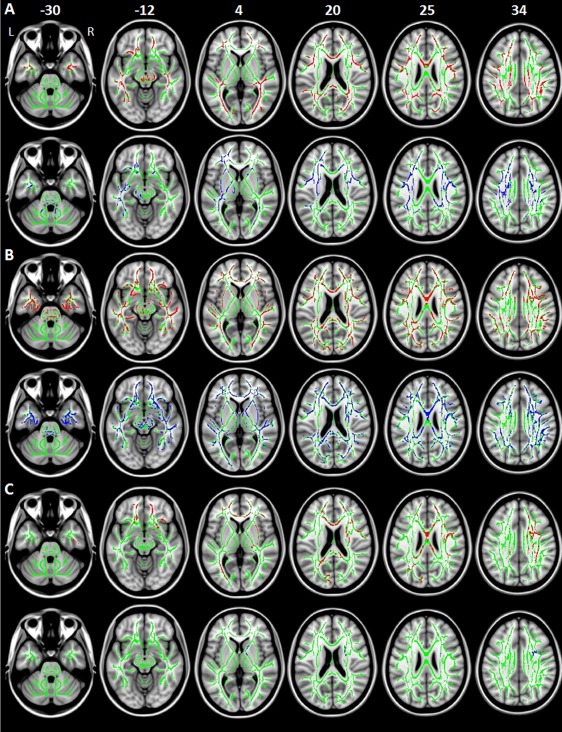
Decreased fractional anisotropy (FA, red) and increased mean diffusivity (MD, blue) in: A) patients with Parkinson's disease and freezing of gait (PD‐FoG) compared with healthy controls; B) PD‐FoG patients from the independent sample compared with matched controls; C) PD patients without FoG (PD‐noFoG) from the independent sample compared with matched controls. Results are overlaid on the axial sections of the Montreal Neurological Institute standard brain in neurological convention (right is right), and displayed at P < 0.05 family‐wise error (FWE) corrected for multiple comparisons at the cluster level using the threshold‐free cluster enhancement option. The white matter skeleton is shown in green.