
FIG. 1.
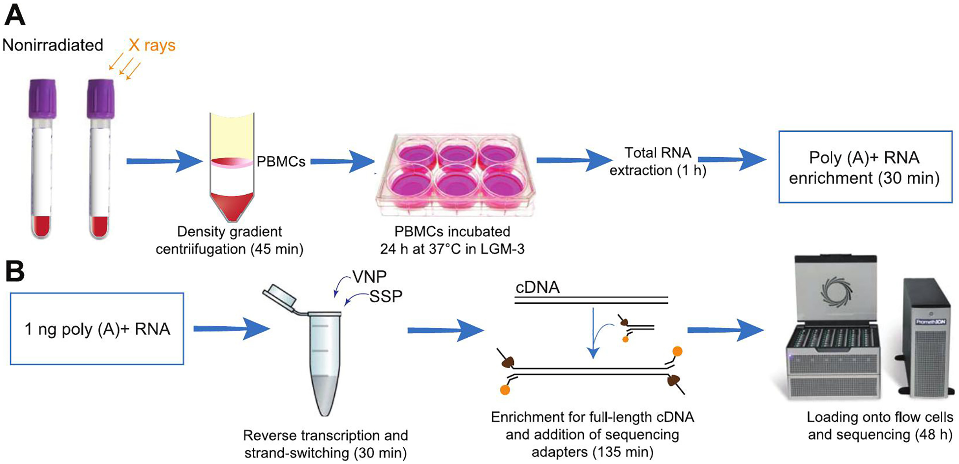
Experimental workflow. Panel A: Blood from nine healthy donors (20 ml per donor), which was sham or 2 Gy irradiated (dose rate 0.5 Gy min−1) ex vivo, was used to isolate PBMCs by a density gradient centrifugation. The PBMCs were incubated for 24 h at 37°C before the RNA was extracted. The total RNA obtained was poly(A)+ enriched to remove ribosomal RNAs from the samples. Panel B: A total of 1 ng from the poly(A)+-enriched RNA was used for the sequencing analysis. The RNA was reverse transcribed using VN and strand-switching (SSP) primers. A PCR amplification step was performed to enrich the samples for full-length cDNAs followed by the addition of sequencing adapters. The samples were run in two SpotON flow cells per sample in a PromethION sequencer. The time required for each step of the protocol is included in the diagram.