
Figure 3.
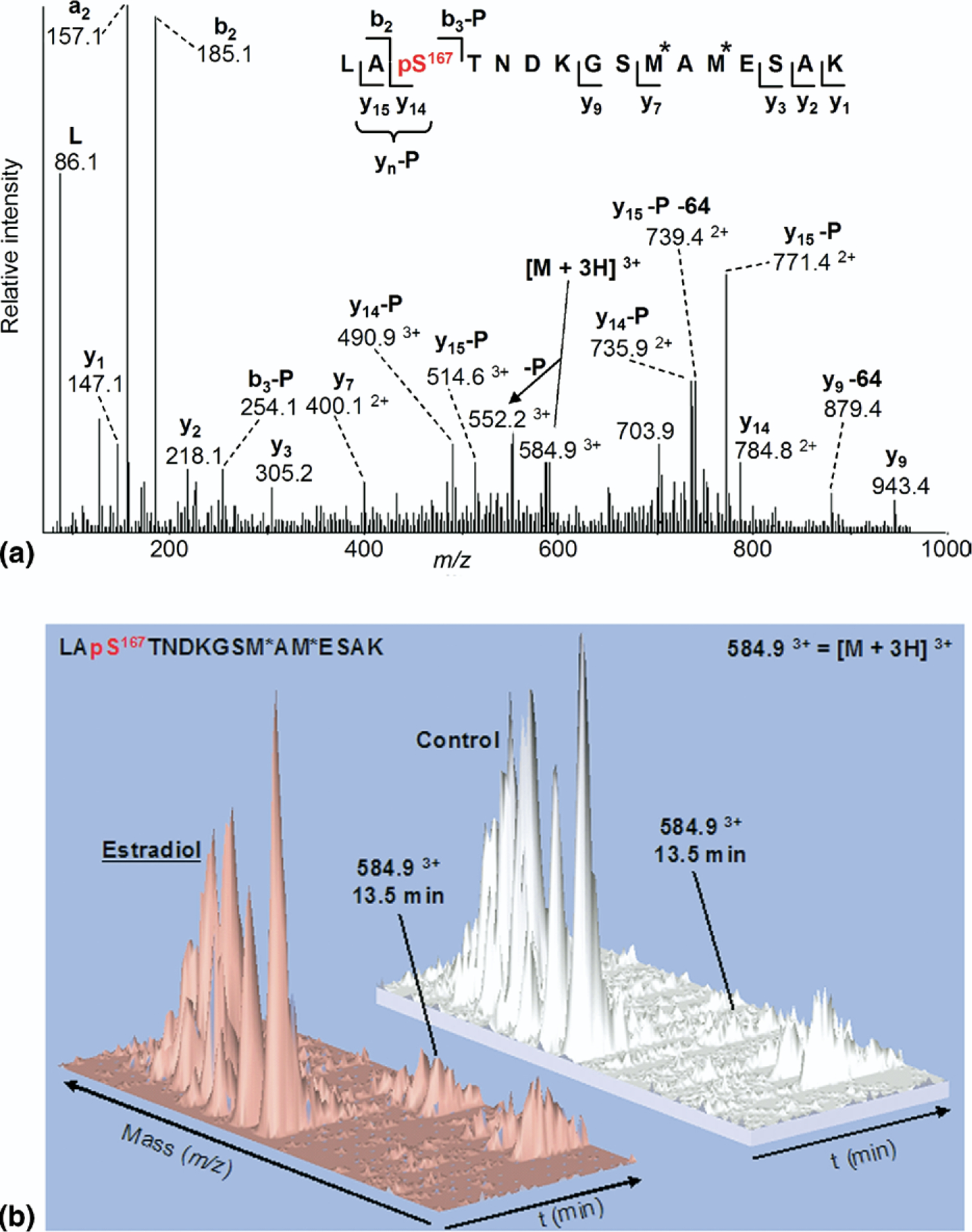
(a) ESI-MS/MS spectrum of triply charged monophosphorylated peptide LApS167TNDKGSM*AM*ESAK (residues 165–180) at m/z 584.9 (M = 1751.72) from tryptic digestion of ERα isolated from MCF-7 cells treated with E2 (M* = mono-oxidized methionine.) (b) three-dimensional plots of ESI-MS representing m/z, retention time, and ion intensity on the x, y, and z axes, respectively, showing increased ion current for phosphorylated peptide m/z = 584.9, eluting at 13.5 min, from cells treated with estradiol compared to untreated control.