
Figure 2.
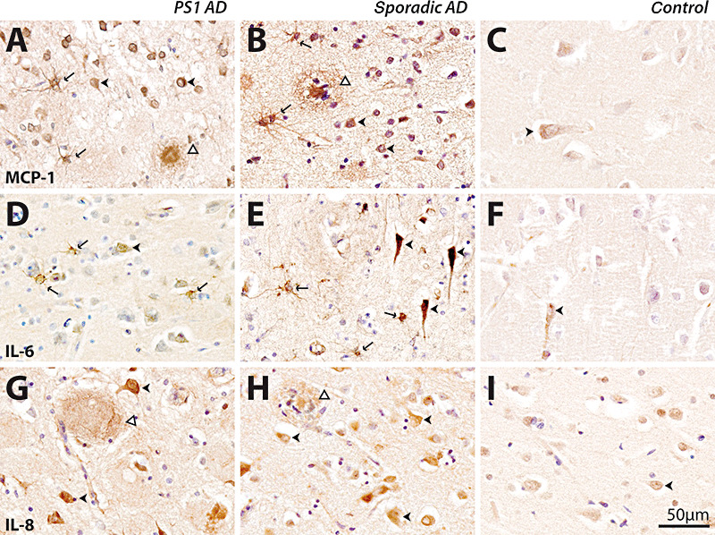
Immunohistochemical detection of monocyte chemoattractant protein‐1 (MCP‐1) (A–C), interleukin‐6 (IL‐6) (D–F) and interleukin‐8 (IL‐8) (G–I) in presenilin‐1 (PS1) AD (A,D,G), sporadic AD (B,E,H) and old controls (C,F,I). Immunoreactivity in young and old controls was similar, so only representative cases from old controls are shown. The scale bar (50 µm) applies to all panels. MCP‐1 immunoreactivity was seen in neurons (arrowheads, A,B), astrocytes (arrows, A,B) and plaques (open arrowheads, A,B) in PS1 (A) and sporadic (B) AD. MCP‐1 immunoreactivity was predominantly observed in neurons in controls (arrowhead, C). Within the cortex IL‐6, immunoreactivity was seen in neurons (arrowheads, D,E) as well as astrocytes (arrows, D,E) in PS1 (D) and sporadic (E) AD. IL‐6 immunoreactivity was predominantly located in neurons in controls (arrowhead, F). IL‐8 immunoreactivity was predominantly seen in plaques (open arrowheads, G,H) and neurons (arrowheads, G,H) in PS1 (G) and sporadic (H) AD. IL‐8 immunoreactivity was predominantly located within neurons in controls (arrowhead, I). AD = Alzheimer's disease.